


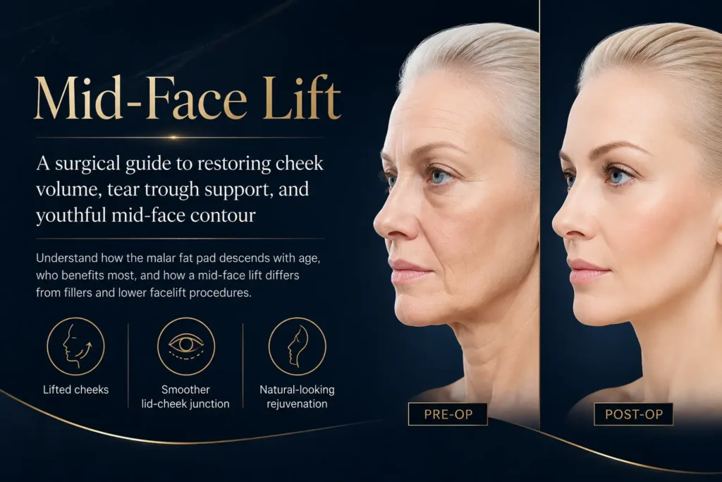
Of all the regions of the face, the mid-face is the one that ages most visibly — and the one most frequently undertreated. A mid-face lift isn’t just about lifting cheeks. It’s about restoring what gravity has displaced: the volume, the contour, and the vitality that defines a youthful face.
When patients describe looking tired even when they feel fine, or seeming sad without reason, or noticing that their under-eye area seems to merge with a flattened cheek — these are almost always mid-face concerns. Not lower face. Not the neck. The middle third of the face: the zone between the lower eyelid and the corner of the mouth. This is the area that transforms a rested, vibrant appearance into one that reads as fatigued or older than it is — and it deserves a targeted surgical response.
This guide covers what a mid-face lift actually involves anatomically, which patients benefit most, the surgical approaches available, recovery expectations, and how it compares to non-surgical alternatives and lower facelift procedures. As always, my goal is to give you the information you’d want from a surgeon who genuinely understands this anatomy — not a brochure.
Understanding Mid-Face Anatomy: What Actually Changes with Age
The mid-face encompasses the region bounded superiorly by the lower eyelid, inferiorly by the upper lip, and laterally by the temporal area. This zone contains multiple discrete fat compartments — including the malar fat pad, the sub-orbicularis oculi fat (SOOF), the nasolabial fat, and the deep medial cheek fat — that descend and atrophy independently at different rates as we age (Rohrich & Pessa, 2007).
The malar fat pad is the key structure. It’s the rounded, full cushion of fatty tissue that sits directly beneath the eye when you’re young — the “apple” of your cheek that catches light and gives the face its characteristic youthful fullness. It sits in the superficial layer of the face, attached at its upper border to the dermis overlying the cheekbone and bound inferiorly by the nasolabial fold.
With age, the malar fat pad doesn’t disappear — it descends. The supporting ligamentous structures that anchor soft tissue to the underlying bone gradually attenuate, and the fat pad shifts downward and inward, away from the orbital rim and toward the nasolabial region. This descent is compounded by bone resorption at the cheek skeleton, which reduces the underlying scaffold; by thinning of the dermis, which reduces the skin’s ability to maintain tissue position; and by volume reduction within the fat compartments themselves as adipose tissue atrophies.
The result is a characteristic triad that patients recognise intuitively even if they can’t name it anatomically: a hollow transition between the lower eyelid and cheek (the tear trough or nasojugal groove), a flattened or sunken mid-cheek, and deepening of the nasolabial folds. Together, these changes create an appearance of fatigue, sadness or premature aging — regardless of how a person actually feels.
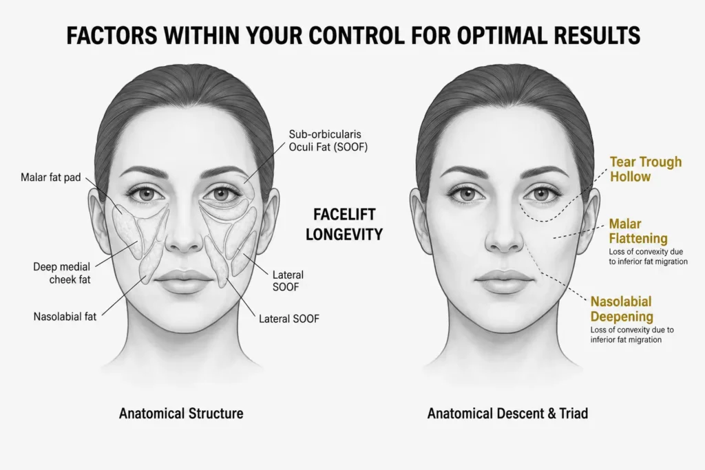
| Change 1
Tear Trough Formation
As the malar fat pad descends away from the orbital rim, it exposes the bony orbital margin and creates a visible hollow — the tear trough — at the lid-cheek junction. This is often the earliest visible sign of mid-face aging. |
Change 2
Malar Flattening
The cheek loses its convex projection as the malar fat pad migrates inferiorly. The face shifts from the inverted-triangle silhouette of youth to a more rectangular, descended shape. |
|
| Change 3
Nasolabial Deepening
As descended tissue stacks above the nasolabial fold, the fold itself deepens and widens. This is a consequence of tissue migration, not primarily of skin laxity — which is why filler alone often fails to fully resolve it. |
Change 4
Malar Bags & Festoons
In some patients, descent of the cheek tissue combined with orbicularis oculi laxity produces bulges beneath the eye — malar bags — that create shadows and a persistently tired appearance. |
What Is a Mid-Face Lift, Specifically?
A mid-face lift — also referred to as a cheek lift, malar lift, or subperiosteal mid-face lift depending on the technique — is a surgical procedure that addresses age-related changes in the central facial third by vertically repositioning descended soft tissue structures back to their original, more superior anatomical position. Unlike a standard lower facelift, which primarily targets jowl laxity, neck skin, and the jawline, the mid-face lift focuses specifically on the malar fat pad, the SOOF, and the soft tissue overlying the cheekbones and the lower eyelid.
The defining principle of the procedure is repositioning rather than removal. The goal is not to excise tissue or stretch skin — it’s to restore descended structures to where they anatomically belong: higher on the facial skeleton, adjacent to the orbital rim, where they create the smooth, convex cheek contour associated with youth. When performed correctly, a mid-face lift does not produce the pulled or tight appearance that older-style facelifts sometimes created, because the vector of correction is vertical — upward toward the cheekbone — rather than lateral toward the ear.
The mid-face is where I spend the most time in consultation, because it’s the most misunderstood zone. Patients come in requesting tear trough filler, or asking why their filler isn’t lasting. Frequently the answer is that filler addresses the shadow but not the structural cause — the descended cheek volume above it. When I reposition that tissue surgically and restore it vertically, the tear trough often resolves without any filler at all. That’s the difference between treating the symptom and treating the anatomy.
— ABmedi Team Clinic Surgical Team, Board-Certified Facial Plastic Surgery
Surgical Approaches: How the Mid-Face Lift Is Performed
There is no single standardised technique for mid-face lifting — the approach is selected based on the patient’s anatomy, degree of descent, associated concerns (such as eyelid laxity), and whether the procedure is being performed alone or in combination with other facial surgery. At ABmedi Team Clinic, we use the approach that best serves each individual’s anatomy. The most established techniques include:
3.1. Subciliary / Transconjunctival Approach
Access is gained through a lower eyelid incision — either just below the lash line (subciliary) or inside the lower eyelid (transconjunctival, leaving no visible external scar). This provides excellent direct visibility of the orbital rim and the SOOF layer. The malar fat pad and SOOF are elevated in the subperiosteal or supraperiosteal plane and secured superiorly using suspension sutures to the periosteum or the arcus marginalis. This approach is particularly useful when the patient also has lower eyelid concerns such as excess fat or mild skin laxity, as both can be addressed simultaneously. The transconjunctival variation avoids any visible scar at the cost of slightly reduced access for more extensive dissection.
3.2. Endoscopic Mid-Face Lift
Small incisions are placed within the temporal hairline, and a fine endoscopic camera is used to visualise the mid-face anatomy. Instruments are passed through these access points to release soft tissue attachments, elevate the malar pad, and secure it in a repositioned location. The significant advantage of this approach is minimal visible scarring — a meaningful consideration for patients who are not ready for or do not require a more extensive procedure. The limitation is that it provides less direct access to the lower eyelid-cheek junction compared to the subciliary approach, and is best suited to patients with early-to-moderate mid-face descent rather than significant ptosis.
3.3. Deep Plane Mid-Face Lift (via Facelift Incision)
When a mid-face lift is performed in conjunction with a comprehensive facelift — which is frequently the case in patients with combined lower-face and mid-face aging — the deep plane approach allows the surgeon to access and reposition the malar fat pad as a continuous composite flap with the SMAS. Ligament release, including the zygomatic ligaments, allows vertical repositioning of the entire mid-face tissue block without skin tension. This is the most comprehensive approach and delivers the most durable results, but it carries a longer recovery and is most appropriate when the lower face and neck also require correction.
3.4. Gingivolabial Sulcus (Intraoral) Approach
An incision inside the upper lip allows access to the anterior mid-face skeleton and soft tissue. This approach leaves absolutely no visible scar and is particularly useful for subperiosteal dissection and malar implant placement if volume augmentation is planned in addition to repositioning. Its limitation is restricted access to the lateral cheek and reduced ability to address the eyelid-cheek junction directly.
Who Benefits Most — and Who Doesn’t
| Strong Candidates | Less Suitable Candidates | |
|
|
It’s worth noting that mid-face lifting is frequently combined with other procedures rather than performed in isolation. Common combinations at ABmedi Team Clinic include: lower eyelid surgery (blepharoplasty) to simultaneously address excess eyelid fat and skin; fat grafting to restore volume in areas of true adipose atrophy; and full facelift (SMAS or deep plane) when lower-face descent is also present. A thorough consultation — including physical examination of tissue mobility, skin quality, fat compartment distribution, and skeletal structure — determines which approach or combination is appropriate.
| 1–2 hrs
Typical operating time (isolated mid-face) |
7–14 days
Social recovery (return to work) |
5–10 yrs
Typical longevity of structural result |
Mid-Face Lift vs. Lower Facelift: Understanding the Difference

| Factor | Mid-Face Lift | Lower Facelift |
| Primary Target Zone | Mid-cheek, malar fat pad, tear trough, nasolabial folds | Jowls, jawline, neck, lower cheek |
| Direction of Lift | Vertical — upward toward the orbital rim | Lateral-oblique — upward and toward the ear |
| Main Incision Options | Subciliary, transconjunctival, temporal, intraoral — often scarless or minimal | Pre- and post-auricular, extending into hairline |
| Addresses Tear Trough | Yes — directly, through orbital rim tissue elevation | No — lower facelift does not address this region |
| Addresses Jowling | Minimally — not the primary application | Yes — this is the core target |
| Addresses Nasolabial Folds | Yes — through tissue repositioning superiorly | Partially — indirect improvement through SMAS elevation |
| Volume Restoration | Yes — repositioning restores apparent cheek volume | Limited — can be supplemented with fat grafting |
| Recovery | 7–14 days social; 4–6 weeks full | 14–21 days social; 6–8 weeks full |
| Often Combined With | Blepharoplasty, fat grafting, brow lift | Neck lift, platysmaplasty, fat grafting |
Non-Surgical Alternatives: When Fillers Are and Aren’t the Answer
The Role of Dermal Fillers
Dermal fillers are effective for restoring volume lost to aging, softening tear troughs, and temporarily projecting the cheekbones. They are ideal for patients with early-to-moderate changes and good skin quality who do not yet have significant tissue descent. Results typically last 12–24 months.
When Fillers Reach Their Limit
If your primary issue is tissue descent (the malar fat pad has moved downward), adding more filler often leads to an overfilled, unnatural appearance. In these cases, you need repositioning, not just more volume. Fillers cannot structurally correct the downward migration of facial tissue.
The “Pinch & Lift” Test
The clearest sign that surgery is the better option: pinch your mid-cheek tissue and lift it slightly toward the eye area. If the appearance improves significantly, your concern is tissue descent, which responds best to a mid-face lift.
A Combined Approach
Fillers and surgery are often complementary. Many patients at ABmedi Team Clinic benefit from a mid-face lift to reposition tissue, supplemented by fat grafting or fillers to refine volume. The goal is to use the right tool for the specific anatomical problem.
Recovery: A Realistic Week-by-Week Timeline
Days 1–2: Initial Healing Expect swelling and bruising around the eyes and upper cheeks. It is essential to keep your head elevated and use cold compresses to manage discomfort. Pain is typically well-controlled with prescribed medication.
Days 3–5: Peak Symptoms Bruising usually peaks and begins to migrate downward while swelling remains significant. Keep physical activity to a minimum, focusing on rest and short, light walks.
Days 6–10: Early Improvement Bruising clears noticeably, and sutures are typically removed between days 5 and 7. You may feel more comfortable in social settings, though some swelling persists in the mid-face.
Weeks 2–4: Returning to Routine Most bruising resolves, allowing many patients to return to work and low-key social activities. The cheeks will appear fuller and higher as residual swelling continues to fade.
Months 2–3: Settling In Deep swelling resolves, and facial sensations return to normal. The improvements to the lid-cheek junction and nasolabial folds become fully visible as the results settle naturally.
6 Months+: Final Result The repositioned tissues have fully integrated, and any incision scars should be faded and discreet. Patients enjoy a refreshed, youthful contour that typically lasts five to ten years.
⚠ Important: Sub-Orbital Swelling Takes Time
- The eyelid-cheek junction is the region that tends to hold swelling the longest — often 8–12 weeks before appearing fully natural
- Do not judge the final result before 3 months; early post-operative photographs are not representative of the outcome
- Sleep elevated for a minimum of 3 weeks; this significantly reduces the duration of periorbital swelling
- Avoid blood thinners, alcohol, and high-sodium diet during recovery — all of these exacerbate and prolong swelling
- Intense sun exposure should be avoided for at least 3 months; UV accelerates scar maturation and pigmentation
Frequently Asked Questions
Q1: Is a mid-face lift the same as a cheek lift?
Yes — these terms are used interchangeably. A cheek lift, malar lift, and mid-face lift all refer to procedures that address the central third of the face by repositioning descended malar soft tissue. The specific technique may vary (endoscopic, subciliary, deep plane) but the anatomical target and goal are the same.
Q2: Can a mid-face lift fix my tear troughs without lower eyelid surgery?
In many cases, yes. When the tear trough is primarily caused by descent of the malar fat pad away from the orbital rim, repositioning that tissue upward through a mid-face lift can significantly improve the lid-cheek junction and reduce the shadow of the tear trough without any direct eyelid intervention. However, when the tear trough is accompanied by excess lower eyelid fat (herniated orbital fat) or significant lower eyelid skin laxity, blepharoplasty is typically recommended in combination. Assessment during consultation determines which applies.
Q3: Will a mid-face lift make me look pulled or unnatural?
Not when performed correctly. The pulled appearance associated with older facelift techniques resulted from lateral skin tension — skin pulled sideways toward the ears. The mid-face lift uses a vertical vector, repositioning tissue upward toward the orbital rim where it originally sat. This direction of correction restores natural anatomy rather than distorting it. The result should look like a younger, more rested version of you — not an operated one.
Q4: How does a mid-face lift compare to dermal fillers for cheek enhancement?
Dermal fillers add volume; a mid-face lift repositions descended tissue. For patients whose primary issue is volume atrophy (fat has genuinely reduced), fillers or fat grafting are appropriate tools. For patients whose issue is tissue descent (fat has moved downward), fillers placed above the fold may partially compensate but do not correct the underlying position. Surgery repositions; fillers supplement. Many patients benefit from both, used for the problems each actually solves.
Q5: Can a mid-face lift be combined with other procedures?
Yes — and it frequently is. Common combinations include lower eyelid blepharoplasty (when excess fat or skin laxity is also present), fat grafting (to restore volume to areas of true atrophy), brow lift (to address upper facial descent simultaneously), and full facelift (when jowling and neck laxity are also concerns). Combining procedures allows a single recovery period and delivers more comprehensive rejuvenation. The appropriateness of combination surgery depends on overall health, operative time, and the specific concerns being addressed.
Q6: How long do mid-face lift results last?
Results from a well-performed mid-face lift typically last five to ten years, though this varies meaningfully depending on technique depth, skin quality, lifestyle factors (sun exposure, smoking, weight fluctuation) and the rate of individual aging. Deeper approaches with subperiosteal dissection generally produce more durable results than more superficial techniques. The procedure does not stop future aging, but it resets the structural position of the mid-face by a meaningful number of years — and non-surgical maintenance afterward (appropriate skincare, selective filler use) can extend the longevity of the surgical result.
📞 💬 WhatsApp / Chat: [http://wa.me/84975142666] 🌐 Book online: [+84-975-142-666]
Consultations are available in-clinic and online for international patients. Most patients leave their first appointment knowing exactly what they need — and feeling confident about the path ahead.
