


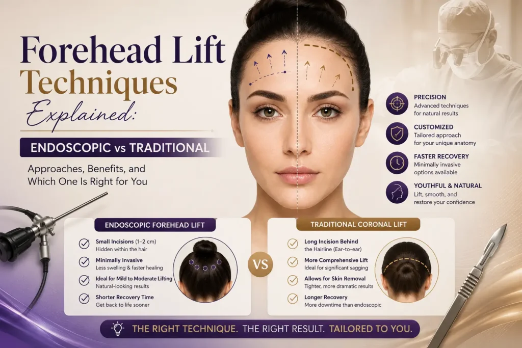
The word “forehead surgery” makes many patients stop cold — imagining a long, visible scar running across their scalp, or a weeks-long recovery that’s hard to hide. Those fears are legitimate, but they belong to a different era of plastic surgery. Here’s exactly how the procedure works today, and which technique is most likely right for you.
How Is a Forehead Lift Performed? (The Step-by-Step Surgical Process)
Before we break down the different techniques, it’s worth understanding what every forehead lift has in common — the fundamental surgical logic that applies whether your surgeon is using an endoscope or a conventional open approach.
| 1 | Anesthesia
Most forehead lifts are performed under local anesthesia combined with IV sedation, meaning you are deeply relaxed and entirely comfortable but do not require general intubation. For more extensive or combined procedures, general anesthesia may be used. Your anesthesia protocol at ABmedi Team Clinic is selected based on the scope of the procedure and your individual health profile. |
| 2 | Incision Placement
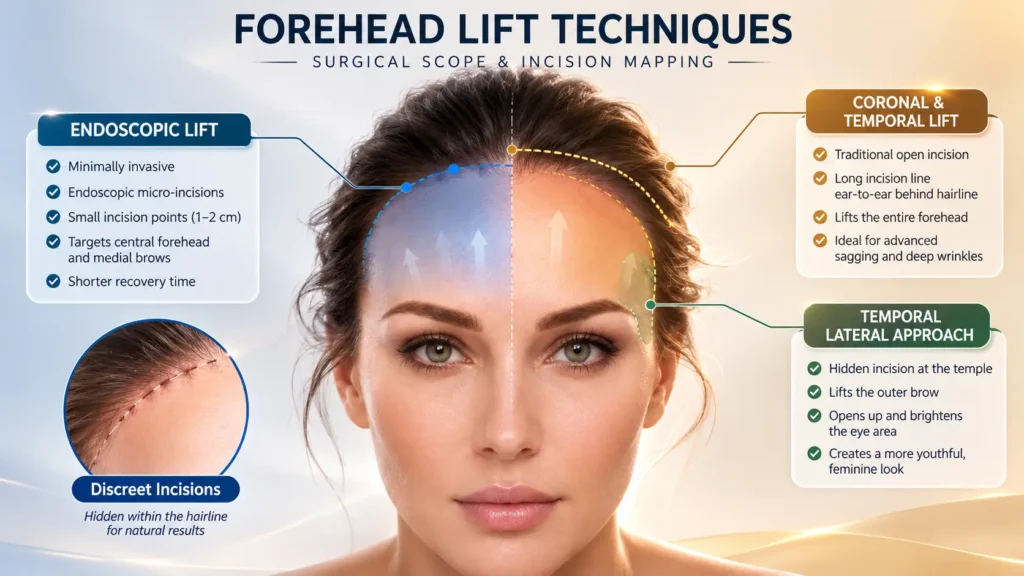
Depending on the chosen technique, the surgeon places one or several carefully designed incisions. These are positioned either within the hair-bearing scalp or immediately at the hairline — always with scar concealment as the governing priority. In an endoscopic approach, three to five short cuts of roughly 1–2 cm each are made; in a coronal lift, a single continuous incision runs the width of the scalp behind the hairline. |
| 3 | Tissue Dissection & Visualization
The surgeon lifts the skin and creates a working plane between the scalp tissue and the skull. In endoscopic surgery, a miniaturized camera (endoscope) is inserted through one of the incisions, projecting a magnified, real-time image of the underlying anatomy onto a monitor. This allows precise visualization of the supraorbital and facial nerves — significantly reducing the risk of inadvertent nerve injury compared to open techniques that rely on tactile feel alone. |
| 4 | Muscle & Tissue Adjustment
The corrugator and procerus muscles — the muscles responsible for frown lines and the “11s” between the brows — are selectively weakened or partially released. The temporalis muscle and temporal fascia are repositioned to elevate the outer brow. This structural adjustment is what distinguishes a forehead lift from surface-level treatments: the correction addresses the underlying cause, not just the visible symptom. |
| 5 | Fixation & Brow Securing
Once the tissues are repositioned to the desired height and tension, fixation sutures or absorbable anchoring devices are placed to hold the brow in its new, elevated position while healing consolidates. The fixation strategy varies by technique and by individual anatomy — your surgeon’s choice here directly influences how long the result holds. |
| 6 | Skin Closure
Excess skin is trimmed where necessary (in open techniques) and incisions are closed with fine sutures or surgical staples. Endoscopic incisions are small enough that closure is minimal. Dressings and a gentle compression wrap are applied. Most patients go home the same day. |
Duration & Setting
A standalone endoscopic or temporal forehead lift typically takes between 45 minutes and 90 minutes. When combined with upper blepharoplasty or a facelift, total operating time increases accordingly. All procedures at ABmedi Team Clinic are performed in a fully accredited surgical suite with anesthesiologist support.
What Are the Different Types of Forehead Lift?
Three primary surgical approaches dominate modern forehead rejuvenation. They share the same goal — an elevated, smoother upper face — but differ significantly in incision design, invasiveness, recovery, and the anatomical problems each is best suited to correct.
What Is an Endoscopic Forehead Lift?
| Endoscopic Forehead Lift | |
| The endoscopic forehead lift is the dominant technique in contemporary facial surgery, now accounting for the majority of brow rejuvenation procedures worldwide. It represents the shift from aggressive open surgery toward precision, camera-guided intervention that delivers equivalent correction with a fraction of the tissue disruption.
The procedure uses three to five small scalp incisions — each typically 1 to 2 centimeters in length — placed strategically within the hair-bearing zone, well behind the hairline. Through these openings, the surgeon passes both the endoscope (camera) and slender operative instruments, working entirely under magnified video guidance. There is no need to create a wide scalp flap or remove large amounts of skin. This approach is particularly effective for patients with mild to moderate brow descent, persistent frown lines, and those who want a natural-looking result with minimal visible evidence of surgery. Because the skin is not removed — tissues are instead repositioned and held with internal fixation — the result is a refreshed elevation rather than a pulled or stretched appearance. |
|
| Incisions | 3–5 cuts, each 1–2 cm, hidden within hair-bearing scalp |
| Invasiveness | Minimally invasive; no large scalp flap created |
| Skin Removal | None — tissue repositioned, not excised |
| Best For | Mild–moderate brow descent; patients with normal-to-low hairline; those prioritizing minimal scarring and faster recovery |
| Limitation | Not ideal for patients with a very long forehead or requiring significant hairline lowering |
What Is a Traditional Coronal Forehead Lift?
| Traditional Coronal (Open) Forehead Lift | |
| The coronal forehead lift is the original “classic” technique, refined over decades before the endoscope changed the field. It involves a single, continuous incision that follows a headphone-shaped arc from approximately ear level, over the crown of the scalp, and down to the opposite ear. The incision is placed several centimeters behind the anterior hairline — ideally deep enough that it sits comfortably within dense hair and is not visible under normal circumstances.
Through this wide access, the surgeon elevates the entire forehead skin as a single continuous flap, gaining excellent visibility of the underlying muscles and allowing for thorough correction of deep furrows, heavy brow descent, and significant forehead laxity. Excess scalp skin is trimmed at the incision point before closure. The trade-off is well-documented: a longer recovery, a more extensive scar (though one concealed within the hair), temporary scalp numbness behind the incision line, and a tendency to elevate the frontal hairline — which makes this approach less suitable for patients who already have a high forehead. Where it remains the technique of choice is in patients with very severe brow ptosis, very thick or heavy forehead tissue, or those who have had prior endoscopic surgery with incomplete correction. |
|
| Incisions | Single continuous cut, ear-to-ear across the scalp crown, several cm behind the hairline |
| Invasiveness | Most invasive of the three primary techniques |
| Skin Removal | Yes — excess scalp skin excised at closure |
| Best For | Severe brow ptosis; deep forehead furrows; thick brow tissue; patients with a low hairline |
| Limitation | Elevates hairline; longer recovery; risk of prolonged scalp numbness posterior to incision |
What Is a Temporal Brow Lift?
| Temporal (Lateral) Brow Lift | |
| The temporal brow lift takes a focused approach: rather than lifting the entire forehead, it addresses exclusively the outer third of the eyebrow — the lateral tail that most commonly descends with age, creating the characteristic downward slope that makes the face look tired or sad at the corners of the eyes.
Two incisions, each roughly 2–3 centimeters in length, are placed within the temporal hairline — one on each side of the head, positioned just above and behind the temple region. Through these access points, the surgeon elevates the skin along the temple and lateral brow, removes a small amount of excess tissue or scalp, and secures the lifted tissue with sutures. The central forehead and the area between the brows are not addressed by this technique. Because only two small incisions are involved and the dissection is localized, a temporal lift can often be performed under local anesthesia alone, with recovery measured in days rather than weeks. It is particularly valued as a complement to upper eyelid surgery — lifting the outer brow while blepharoplasty addresses the lid — and as an adjunct to injectable treatments that handle the central forehead. |
|
| Incisions | Two incisions, 2–3 cm each, within the temporal hairline on each side |
| Invasiveness | Least invasive of the three surgical techniques |
| Skin Removal | Outer one-third of brow and temple only; central forehead unchanged |
| Best For | Isolated lateral brow descent; patients combining with blepharoplasty; those needing subtle targeted correction |
| Limitation | Does not address frown lines, central brow depression, or forehead wrinkles |
Which Forehead Lift Technique Is Best for You?
The “best” technique is not universal — it is the one aligned with your specific anatomy, the degree of correction required, your hairline position, and your recovery constraints. The table below distills the clinical decision-making framework used at ABmedi Team Clinic.
| Criteria | Endoscopic Lift | Coronal (Open) Lift | Temporal Lift |
| Incision Size | 3–5 cuts, each ~1–2 cm
Minimal |
One continuous cut, ~25–30 cm ear-to-ear
Extensive |
2 cuts, ~2–3 cm each at temples
Very small |
| Invasiveness | Minimally invasive | Most invasive | Least invasive |
| Recovery Time | 1–2 weeks to return to social activities; bruising and swelling largely resolving by 10–14 days | 2–4 weeks for initial recovery; full resolution 4–6 weeks | 3–7 days for most patients; stitches dissolve, no removal needed |
| Longevity of Results | Long-lasting; comparable to coronal at 1-year follow-up; gradual natural aging continues
Excellent |
Durable initially; evidence of gradual brow position drop over 5 years in some cases
Excellent |
Good; localized correction holds well; central brow may still descend over time
Good |
| Scarring Risk | Minimal; multiple small scars hidden entirely within dense hair
Very low |
Longer scar hidden within hair; risk of numbness posterior to incision; potential hairline elevation
Moderate |
Two discreet temporal scars; well-hidden; dissolving sutures used
Very low |
| Hairline Effect | Neutral — no hairline elevation | Elevates hairline; unsuitable for patients with high foreheads | Neutral in the central zone |
| Correction Scope | Full forehead: central brow, frown lines, lateral brow | Full forehead: most comprehensive single-procedure correction | Lateral brow and temple only |
| Ideal Patient | Mild–moderate brow descent; normal or low hairline; prioritizing minimal downtime | Severe brow ptosis; deep furrows; thick tissue; low or normal hairline | Isolated outer brow drooping; often combined with blepharoplasty or injectables |
The decision framework at ABmedi Team Clinic follows a clear logic: we start with the least invasive technique that achieves the necessary correction, and escalate only when anatomy demands it. For most patients presenting in their 40s and 50s with mild-to-moderate brow descent and a normal hairline, the endoscopic approach delivers results that are clinically equivalent to open surgery — with a significantly shorter recovery and lower scar burden.
At a glance — which technique fits your situation:
| Endoscopic
You’re likely a candidate if…
|
Coronal (Open)
You’re likely a candidate if…
|
Temporal
You’re likely a candidate if…
|
Scar Management: Where Are the Incisions Placed?
The question patients most often arrive with — and rightly so — is: “Will anyone be able to see the scar?” In the hands of an experienced surgeon using modern technique, the honest answer for the vast majority of patients is no. Here is exactly where each incision goes.
| Endoscopic Forehead Lift — Incision Mapping |
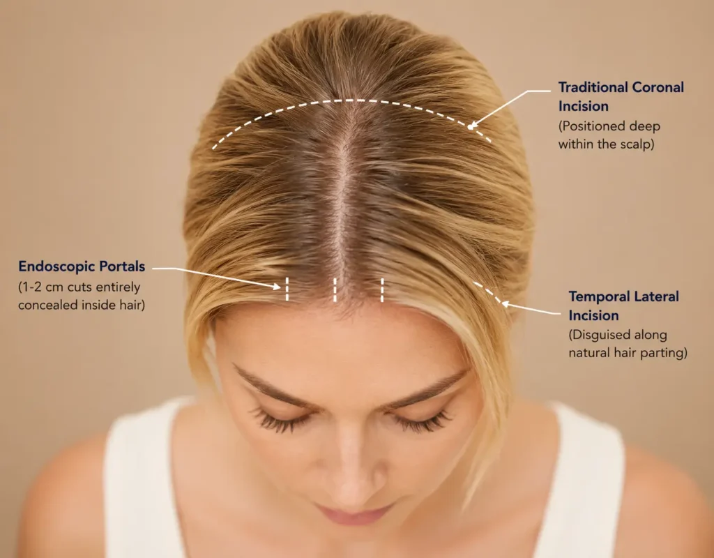
| Three to five incisions are placed in a distributed pattern across the scalp, each positioned approximately 2–4 centimeters behind the anterior hairline within the hair-bearing zone. The exact locations typically include two paramedian incisions (near the crown, on either side of the midline), one or two temporal incisions (in the temporal hair region), and occasionally a midline incision depending on the extent of central brow work required.
Each cut is roughly 1 to 2 centimeters long — about the length of a fingernail. They sit well within the scalp’s hair-bearing tissue, meaning even closely cropped hair will cover them. There is no linear scar, no continuous line that could catch the eye; just a handful of small, scattered points that become progressively less visible as hair regrows around them over four to eight weeks. Length: 1–2 cm per incision Placement: 2–4 cm behind hairline Number: 3–5 incisions Visibility: None under normal hair |
| Coronal (Open) Forehead Lift — Incision Mapping |
| The coronal incision is a single, continuous cut running from approximately the level of the upper ear on one side, arcing over the crown of the head, and descending to the same level on the opposite side — often described as following the path of a wide headphone band. The incision is placed approximately 4–6 centimeters behind the anterior hairline, deep within the hair-bearing scalp.
Because the cut runs through dense hair-bearing tissue, the resulting scar is covered naturally by surrounding hair from above. However, it is a longer scar than any minimally invasive approach — roughly 25 to 30 centimeters in total length — and it does carry a risk of a thin band of numbness in the scalp tissue posterior to the incision line, caused by the sensory nerves that are divided in the process. This numbness typically resolves partially over many months, but may be permanent in a small percentage of patients. The hairline will also shift slightly upward with skin removal, which is factored into the pre-operative planning discussion. For patients with a low hairline who want to simultaneously raise the hairline, a modified coronal incision placed exactly at the hairline junction (pretrichial approach) allows for hairline lowering while the brow is lifted — turning a limitation into an additional aesthetic benefit. Length: ~25–30 cm total Placement: 4–6 cm behind hairline Hairline effect: Elevates ~1–2 cm Numbness risk: Posterior scalp, usually temporary |
| Temporal Brow Lift — Incision Mapping |
| Two separate incisions, one on each side of the head, are placed within the temporal hairline — the hair-bearing zone just above and slightly behind the temple, roughly in line with the outer corner of each eyebrow. Each incision runs approximately 2 to 3 centimeters in length, oriented vertically or at a gentle angle to follow the natural hair parting direction, minimizing any disruption to hair follicles along the cut edge.
These incisions are among the most easily concealed in all of facial surgery. Even patients with fine or thinning hair at the temples generally find that the scar is indistinguishable from a natural part line once healed. Sutures used for closure are typically the dissolving variety — no removal appointment needed. Length: 2–3 cm per side Placement: Within temporal hairline, bilateral Sutures: Dissolving — no removal needed Visibility: Effectively zero in most patients |
ABmedi Team Clinic’s Approach to Scar Prevention
Scar quality is determined as much by technique and aftercare as by incision placement. At ABmedi Team Clinic, incisions are closed in layers to minimize surface tension, and patients receive a structured scar management protocol beginning at suture removal — including silicone therapy and UV protection guidance. The vast majority of our forehead lift patients report that by three months post-operatively, their incision sites are undetectable even on close inspection.
Frequently Asked Questions
Q1: Is the endoscopic forehead lift as effective as the traditional open method?
Clinical research confirms that both techniques achieve comparable degrees of brow elevation at one-year follow-up. The endoscopic method does not remove skin — it repositions tissue internally — which means patients with very significant skin excess may require an open approach for optimal correction. For the majority of candidates, however, endoscopic results are equivalent in quality and more favorable in terms of recovery and scarring.
Q2: Will I lose hair around the incision sites?
Temporary thinning of hair immediately adjacent to a scalp incision is possible and is typically due to the surgical trauma to follicles along the cut edge. In the vast majority of cases, full regrowth occurs within three to six months. Permanent hair loss along a forehead lift incision is uncommon when proper technique is used and is more associated with poorly placed or excessively tense coronal incisions.
Q3: How long does the result of a forehead lift actually last?
A surgical forehead lift produces long-lasting structural correction — most patients retain meaningful improvement for ten years or more. You will continue to age after the procedure, but from a significantly elevated baseline. Some studies have noted gradual brow descent in coronal lift patients over a five-year window; endoscopic fixation techniques have evolved specifically to address long-term maintenance of the elevated position.
Q4: Can I combine a forehead lift with other procedures?
Yes, and combination surgery is both common and clinically sensible. The forehead lift is frequently paired with upper eyelid surgery (blepharoplasty) to address both the brow and the lid in one surgical session, or with a facelift to rejuvenate the full face comprehensively. Performing related procedures simultaneously means a single anesthesia and recovery period — which most patients strongly prefer.
Q5: What happens if I don’t have enough hair to cover the incisions?
Patients with thinning hair or receding hairlines require a technique-specific approach. For these individuals, a standard coronal incision would produce a scar that may eventually be visible as hair density decreases further. The preferred alternatives are either an endoscopic approach (with its multiple small, separated incision points that remain non-continuous even as hair thins) or, in some cases, a direct brow or pretrichial incision placed in a forehead crease where it can blend naturally even without hair coverage.
Q6: When can I return to work and exercise after a forehead lift?
For patients undergoing endoscopic or temporal lifts, most are comfortable returning to desk-based work and light social activities within seven to ten days, once initial bruising and swelling have settled to a level that doesn’t obviously signal recent surgery. Strenuous exercise, heavy lifting, and activities that significantly raise blood pressure or heart rate should be avoided for three to four weeks to protect the fixation and support optimal healing.
📞 💬 WhatsApp / Chat: [http://wa.me/84975142666] 🌐 Book online: [+84-975-142-666]
Consultations are available in-clinic and online for international patients. Most patients leave their first appointment knowing exactly what they need — and feeling confident about the path ahead.
